
Color Blindness Test
Test for red-green and blue-yellow color vision deficiencies.
Tip: Please ensure Night Shift, f.lux, or blue-light filter glasses are temporarily paused for accurate chromatic results.
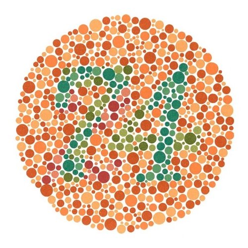
Session Results
Congratulations! Your color vision appears to be normal. You correctly identified most or all of the Ishihara plates.
Statistics
This chart displays global color discrimination accuracy across Ishihara plates.
About Colour Blindness
What it measures
This test evaluates your cone photoreceptor sensitivity and chromatic contrast discrimination across visible wavelengths.
How it works
Each plate uses pseudo-isochromatic dots with randomized luminance and size along clinical color confusion axes. the data
Cone photoreceptors
Human color vision relies on three types of retinal cone cells: L-cones (red), M-cones (green), and S-cones (blue).
Red-Green deficiency
Deuteranomaly (green-weak) and Protanomaly (red-weak) are X-linked traits affecting roughly 8% of biological males.
Blue-Yellow & rare forms
Tritanopia affects short-wavelength S-cones, while Achromatopsia is complete inability to perceive chromatic differences.
Other vision tools
Challenge your executive color interference in the Stroop Effect test. Stroop Test
Colour Blindness & Ishihara Plates FAQ
Color Discrimination & Photoreceptor Sensitivity4 Detailed GuidesColor vision deficiency, Ishihara tests, and prevalence benchmarks.